New method to screen embryos could improve IVF success rates
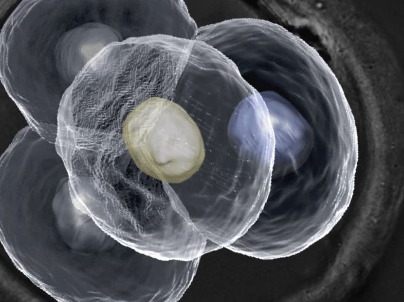
SINGAPORE — Researchers at A*STAR’s Institute of Molecular and Cell Biology (IMCB) have developed new advanced microscopy technologies that will allow the development of an embryo to be monitored in real time without affecting its future growth. Currently, embryos are studied through invasive methods, which do not keep them alive.
SINGAPORE — Researchers at A*STAR’s Institute of Molecular and Cell Biology (IMCB) have developed new advanced microscopy technologies that will allow the development of an embryo to be monitored in real time without affecting its future growth. Currently, embryos are studied through invasive methods, which do not keep them alive.
The technologies have the potential to improve the efficiency of assisted reproduction therapies, the institute said in a statement on Friday (May 20).
“Our lab is the only one in the world imaging single cells in live mammalian embryos at the quantitative level, which allows us to observe every cell within an embryo at every stage of its development,” said Dr Nicolas Plachta, senior principal investigator at IMCB, in the statement.
“Our findings as a result of this advanced technique have put forth a new paradigm of knowledge that would encourage more detailed microscopic analysis for future assisted reproduction procedures.”
Traditional methods of reproduction therapy — such as In-Vitro Fertilisation (IVF) and Preimplantation Genetic Diagnosis — assume that every cell within an embryo is identical prior to implantation.
Using advanced real-time imaging techniques, researchers at IMCB have found that the cells are actually differentiated, with each playing different roles in later development. They reached this conclusion after observing the cells of a preimplantation mouse embryo, which the IMCB says strongly resembles human embryos in their early stages of development.
As these technologies develop further, it could allow fertility specialists to better study the microscopic properties of embryos and decide more accurately if an embryo is suitable for implantation. They will also be able to screen an embryo for genetic abnormalities using imaging lasers instead of physical manipulation.
Noted Dr Sadhana Nadarajah, the director of KK Women’s and Children’s Hospital’s IVF centre: “If (the method) can be successfully used on human embryos, without affecting its successive growth, it will improve the technique of embryo selection in IVF.”
The study was published earlier this year in the scientific journal, Cell.
According to IMCB, more Singaporean women are turning to assisted reproduction therapies. Last year, more than 6,000 of such procedures were carried out in the Republic, compared to less than 5,000 in 2012.


